The left humerus (os brachii)* is seen from the posterior or dorsal surface
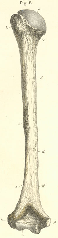
a) Head of the humerus.
b) Greater tubercle of humerus.
c) Neck of the humerus (insertion of capsular ligament).
d) Body of the humerus* diaphysis.
e) Groove for radial nerve.
f) Margin of medial angle (origin for m. triceps* medial head).
g) Medial condyle (flexor) (with groove for ulnar nerve and origin for mm pronator teres* flexor carpi radialis and ulnaris* palmaris longus* flexor digitorum superficialis and ligament laterale internum).
h) Lateral condyle (extensor) (origin for mm triceps* lateral head* supinator longus and brevis* extensor carpi radialis and brevis* extensors digitorum and for ligament laterale externum).
i) Trochlea of humerus.
k) Olecranon fossa.