


 04-14-2012, 10:08 PM
|
رقم المشاركة : 1 | ||||
|
**حصريا لمنتديات عجور**
|
||||

|


|
04-14-2012, 10:20 PM
|
رقم المشاركة : 2 | ||||
|
|
||||
|
|
|
04-14-2012, 10:43 PM
|
رقم المشاركة : 3 | ||||
|
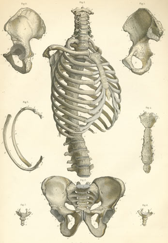
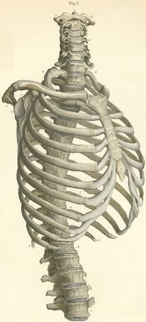
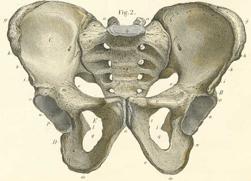
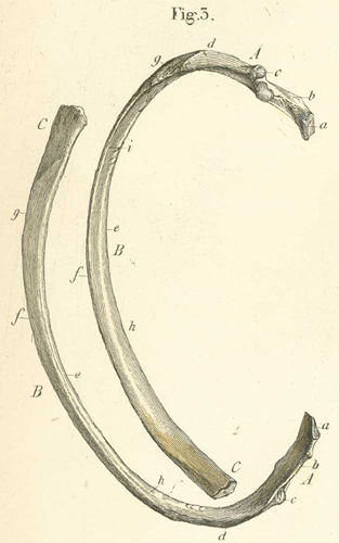
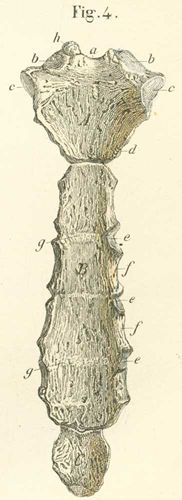
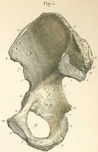
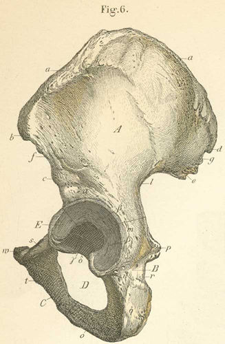
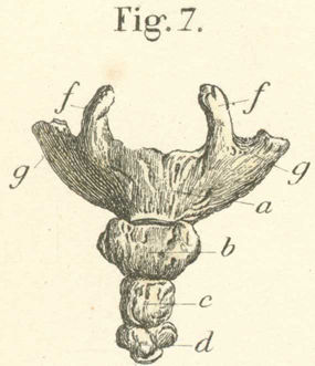
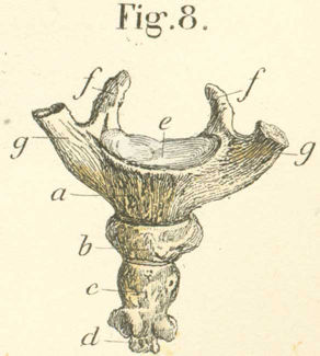
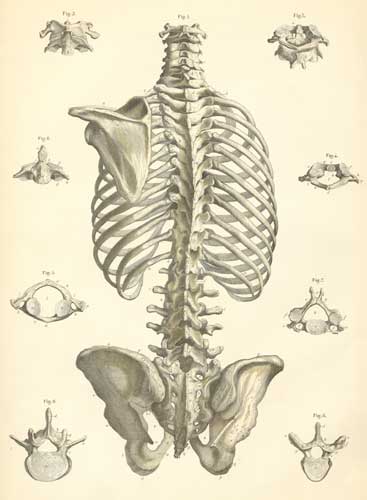
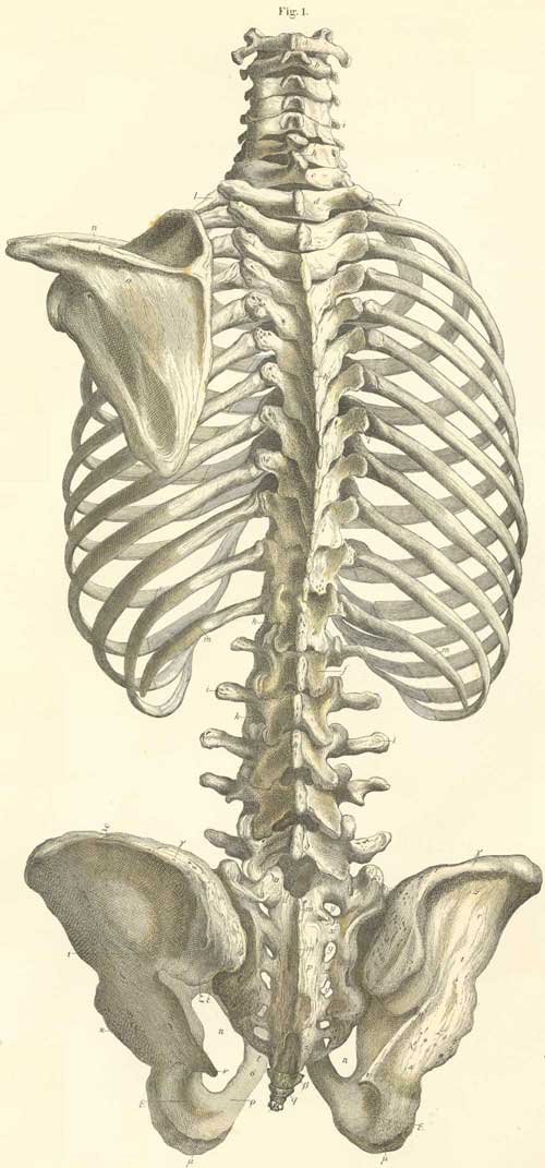
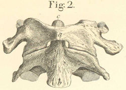
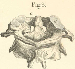
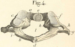
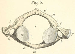
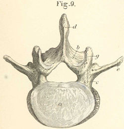
Bones of the trunk |
||||
|
|
|
04-14-2012, 10:54 PM
|
رقم المشاركة : 4 | ||||
|
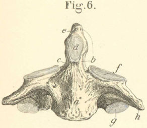
Bones of the trunk |
||||
|
|
|
04-15-2012, 12:44 AM
|
رقم المشاركة : 6 | ||||
|
يعطيك العافية ابو اوس على هذا المجهود |
||||
|
|

|
04-16-2012, 12:09 AM
|
رقم المشاركة : 7 | ||||||
|
|
||||||
|
|
|
04-16-2012, 12:11 AM
|
رقم المشاركة : 8 | ||||
|
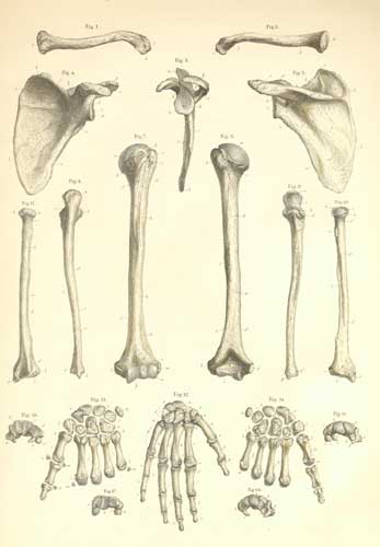
Bones of the upper limb |
||||
|
|
|
04-16-2012, 12:12 AM
|
رقم المشاركة : 9 | ||||
|
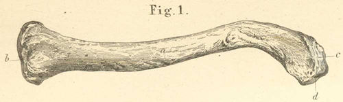
The left clavicle* showing its inferior and posterior surface |
||||
|
|
|
04-16-2012, 12:14 AM
|
رقم المشاركة : 10 | ||||
|
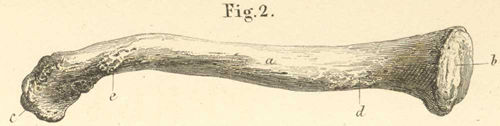
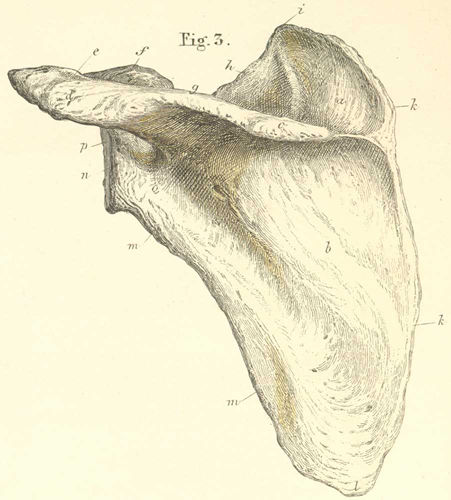
The left scapula* from its posterior or outer surface |
||||
|
|
 |
| الكلمات الدلالية (Tags) |
| anatomy, منتديات عجور, blood, bones, brain, تمريض, تسريح, تشريح الجسم البشرى, تشريح جسم الانسان, جسم الانسان, muscle, nerve, organs, sex, skull, smell, spinal cord, teeth, علمي, عجور, veins, visual, نبيل زبن, طب |
| الذين يشاهدون محتوى الموضوع الآن : 2 ( الأعضاء 0 والزوار 2) | |
 العرض العادي

|
|
